
Figure 7.
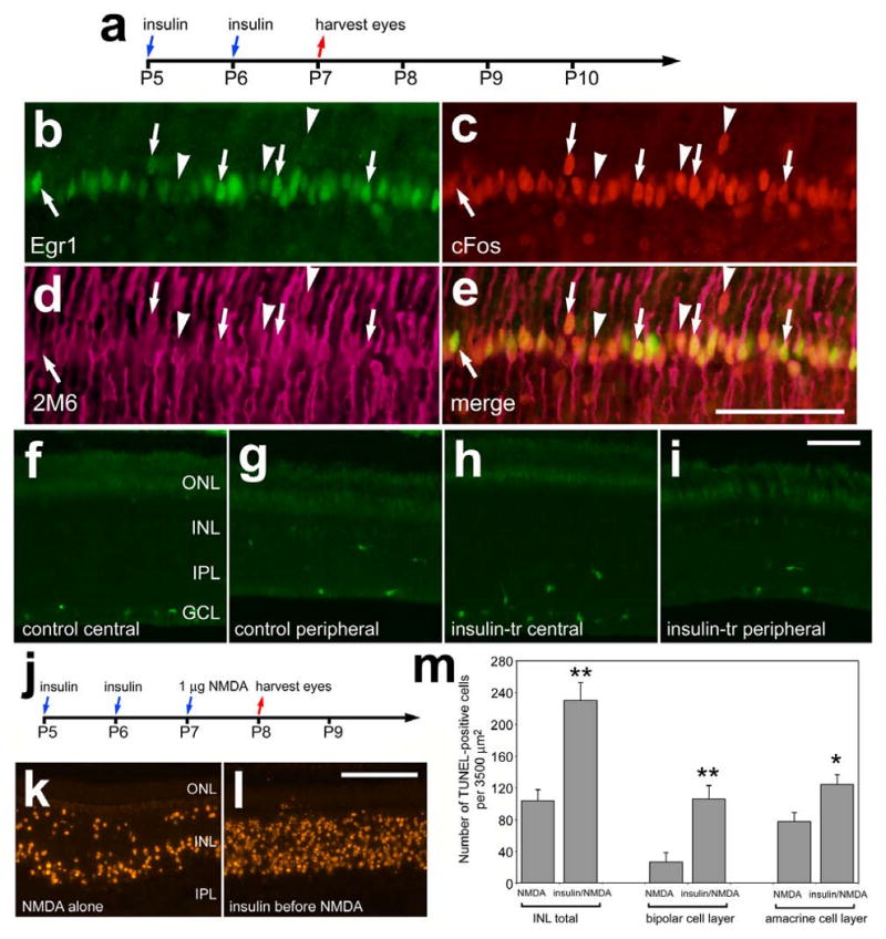
Consecutive daily injections of insulin stimulate the expression of cFos and Egr1, whereas p38 MAPK appear unaffected, in Müller glia and decreases neuronal survival following an excitotoxic insult. Retinas were obtained from eyes that received intraocular injections of 1 μg insulin at P5 and P6, and were harvested at P7 (a-i) or injected with 1 nmol NMDA at P7 and harvested at P8 (j-m). Vertical sections of the retina were labeled with antibodies to Egr1 (green; b and e), cFos (red; c and e), the glial marker 2M6 (magenta; d and e), and p38 MAPK (f-i). Arrows indicate Müller glia nuclei that are immunoreactive for Egr1 and cFos, and arrow-heads indicate Müller glia nuclei that are immunoreactive for cFos alone. Nuclei of cells that contained fragmented DNA were detected by using the TUNEL method (k and l). The histogram in panel m illustrates the mean and standard deviation of TUNEL-positive cells in the distal INL (presumptive horizontal and bipolar cells), proximal INL (presumptive amacrine cells), and total cells in the INL. Significance of difference (*p<0.01, **p<0.0001) was determined by using a two-tailed, unpaired Student's t-test. Abbreviations: ONL – outer nuclear layer, INL – inner nuclear layer, IPL – inner plexiform layer, GCL – ganglion cell layer.