
Figure 8.
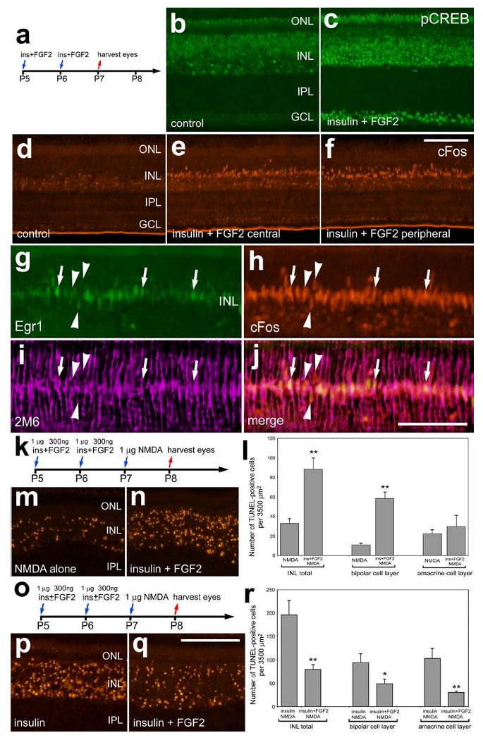
Insulin and FGF2 stimulate glial accumulation of cFos, Egr1 and pCREB and decrease neuronal survival in response to NMDA-treatment. Retinas were obtained from eyes that were injected at P5 and P6 with the combination of insulin and FGF2 or saline and harvested at P7 (a-j), or from eyes that were injected at P5 and P6 with the saline, insulin alone, or the combination of insulin and FGF2, 1 μmol NMDA at P7 and harvested at P8 (k-r). Vertical sections of the retina were labeled with antibodies to pCREB (b and c), cFos (red; d-f and h), Egr1 (green; g) or 2M6 (magenta; i). Arrows indicate the nuclei of 2M6-positive Müller glia that were immunoreactive for Egr1 and cFos, and arrow-head indicate the nuclei of Müller glia that were immunoreactive for cFos alone. The TUNEL method was used to label dying cells (m,n,p and q). The histograms in panels l and r illustrate numbers of dying cells that were increased in retinas treated with the combination of insulin and FGF2 before NMDA, whereas FGF2 counteracts the death-inducing affects of insulin-treatment prior to NMDA. Significance of difference (*p<0.004, **p<0.0001) was determined used a student's t-test. The calibration bar (50 μm) in panel c applies to panels b and c, the bar in j applies to d-j, and the bar in q applies to m, n, p and q. Abbreviations: ONL – outer nuclear layer, INL – inner nuclear layer, IPL – inner plexiform layer, GCL – ganglion cell layer.