
Fig. 8.
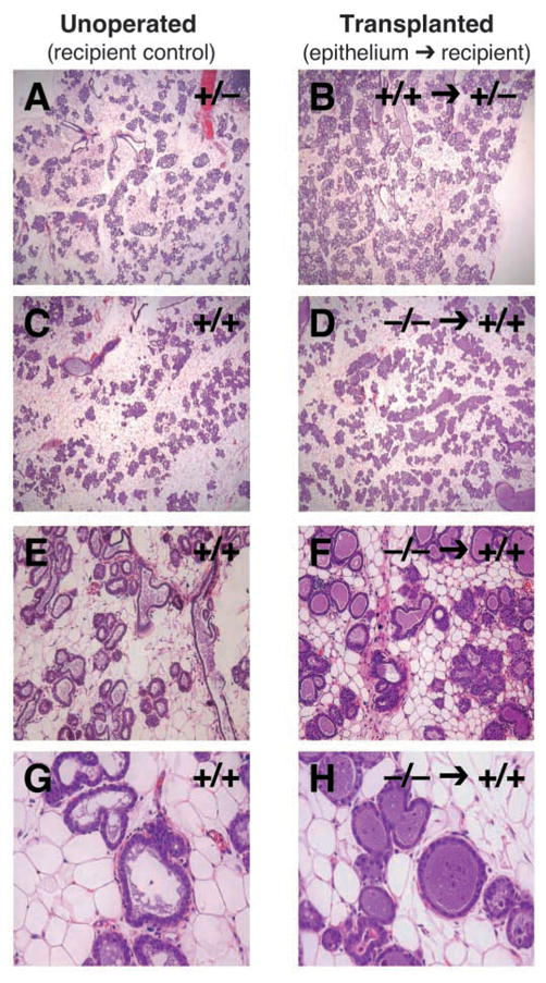
Transplantation of Dgat1−/− epithelium into Dgat1+/+ recipient mice. (A–H) After mammary epithelial transplantation, recipient mice were mated and epithelial morphology was analyzed by histology at L1. Shown are recipient (unoperated) control glands (A,C,E,G) and recipient glands that received transplanted epithelium (B,D,F,H). Control transplantations are shown for comparison (A,B).