
Fig. 1.
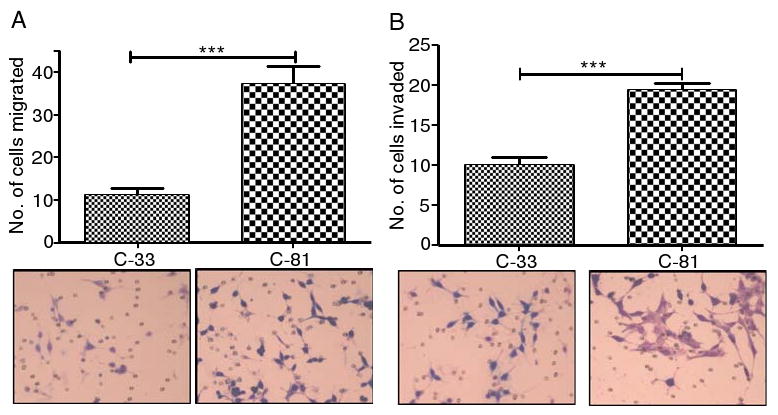
Migration and invasion analysis of LNCaP cells. C-81 and C-33 LNCaP cells of 5 × 10⁴ cells/ml medium were inoculated in the upper chambers of 24-well migration and matrigel invasion chamber plates. After 22–24 h, the cells that transversed to bottom side of the chamber plates were fixed, stained, and then counted under a phase contrast microscope (40×) in three independent fields for each insert. C-81 cells showed highly significant level of migration (a) and invasion (b) than C-33 cells through polyethylene terapthalate membrane and matrigel invasion chambers, respectively. The data, which were expressed as mean±SD, were obtained from three independent experiments (**p<0.05 and ***p<0.001)