
Figure 1.
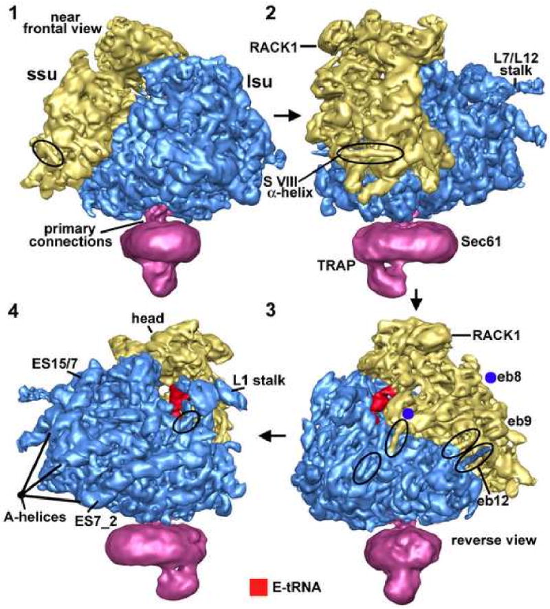
Surface views of the ribosome-channel complex.
In panels 1-4, a rotation series is shown about a vertical axis for the ribosome-channel complex with the ribosome filtered at 8.7Å resolution. The small subunit is shown in gold, the large subunit in blue and the native channel in magenta. Certain key features are indicated. In particular, some surface α-helices are circled and A-form helices of expansion segment RNAs are marked. The E-site tRNA (red) is visible in panels 3 and 4 between the small and large subunits.