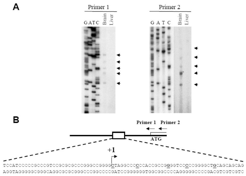
Figure 1. Multiple transcription start-sites upstream of the p39 ATG.
(A) The results of primer extension analysis with primers that hybridize across (Primer 1) or downstream (Primer 2) of the p39 ATG using total RNA isolated from 15 day old mouse brain or liver. Start-sites 63–95bp upstream of the ATG are indicated by arrowheads. DNA sequencing of p39 genomic DNA was performed with the same primers. (B) Schematic of the p39 promoter indicating the positions of primers used in the primer extension reactions. The 5 identified transcription start sites are indicated in bold and underlined. The most 5′ start site relative to the ATG is indicated by the arrow and the nucleotide is assigned +1.

