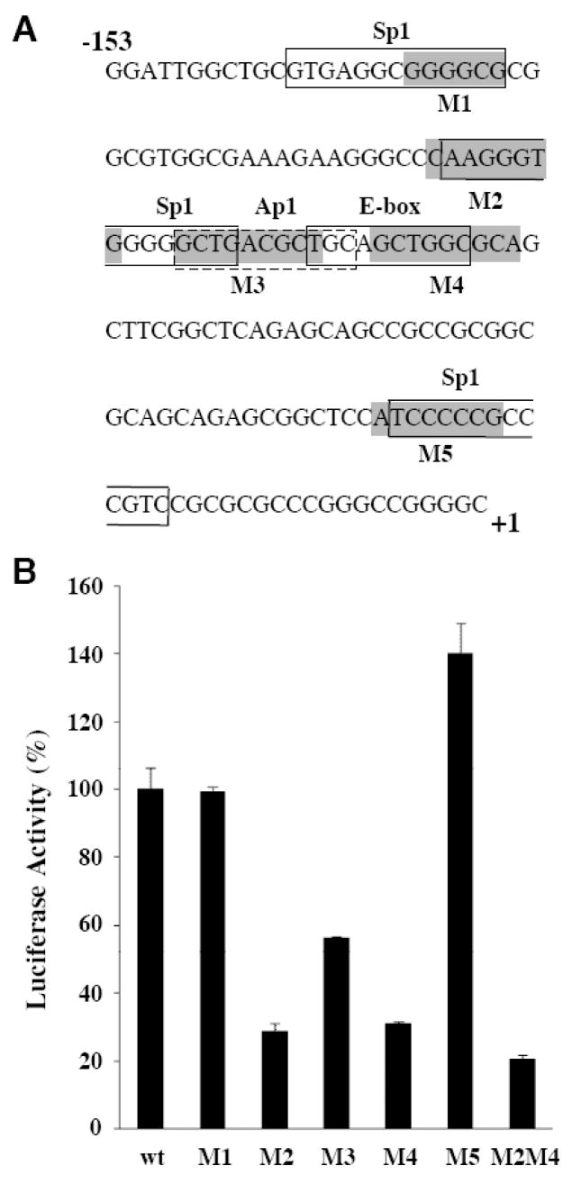
Figure 4. Mutation of the Sp1, AP1/CREB/ATF and E box clustered binding sites reduced the activity of the p39 promoter.
(A) The positions of 6–10bp scanning mutations (M1–M5) in the p39 promoter region are indicated by shaded boxes. Predicted transcription factor binding sites for Sp1, AP-1/CREB/ATF and E box-binding proteins are indicated. (B) Reporter constructs containing the indicated scanning mutations in the p39 promoter construct p39-153/+46 were transfected into N2A cells and the resulting luciferase activity determined. Luciferase activity of the wild-type p39-153/+46 construct was set at 100. The experiments were performed in triplicate; error bars indicate the S.D..

