
Fig. 4.
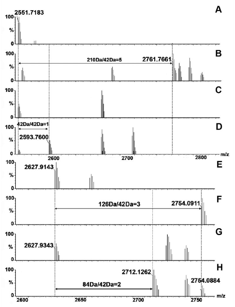
MALDI spectra of peptide fragment NKPAAKTDATIKKEQKLIQAQNL (95-117, m/z 2551, A-D) and fragment FEKTHTVSAHRKAQKAVNLVSFE (120-143, m/z 2627, E-H) of Efb(-C) derived from samples A) free Efb-C without lysine acetylation; B) free Efb-C with lysine acetylation; C) Efb-C/C3d without lysine acetylation; and D) Efb-C/C3d with lysine acetylation. The mass differential of 168 Da (210 Da - 42 Da) in the free protein versus the protein complex indicated that 4 lysine residues in Efb-C were protected upon Efb-C/C3d binding; E) free Efb-C without lysine acetylation; F) free Efb-C with lysine acetylation; G) Efb-C /C3d without lysine acetylation; and H) Efb-C/C3d with lysine acetylation. The mass differential of 42 Da (136 Da - 84 Da) in the free protein versus the protein complex indicated that a single lysine residue in Efb-C was protected upon Efb-C/C3d binding.