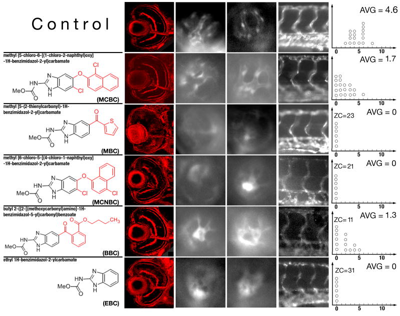
Fig. 5.
Phenotypic changes produced by compounds structurally similar to albendazole and mebendazole. The left-most column of panels shows confocal images of transverse sections through control and chemically-treated retinae. The concentrations of chemicals used for experiments shown in these panels are indicated in Table 2 in italics. Panels in the middle two columns of images show lateral views of the retinal vasculature through the lens at 96 hpf. The right-most column of images shows lateral views of the trunk vasculature. Graphs to the right are plotted as in Fig. 4. In all images, dorsal is up and anterior is left.

