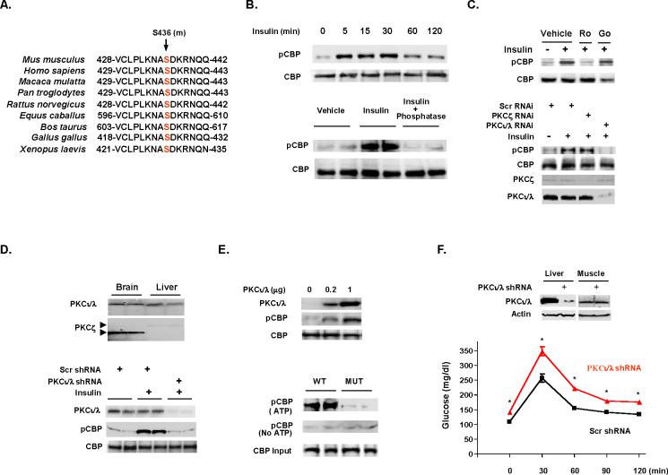
Figure 2.
Insulin induces CBP phosphorylation through aPKC. (A) CBP phosphorylation site at Ser436 (mouse) is conserved across eukaryotic species. (B) Insulin induces CBP phosphorylation in a time-dependent manner in H4IIEC3 cells (top). Hepatic CBP phosphorylation is detected after insulin administration in fasted mice and alkaline phosphatase treatment of hepatic lysates removes phosphorylation (bottom). Each lane represents an individual mouse sample. (C) Nonisoform selective PKC inhibitor Ro31-8220 (Ro), but not Go6976 (Go; inhibitor of classical and novel PKC isoforms) inhibits insulin stimulated CBP phosphorylation (top). RNAi mediated aPKCι/λ knockdown blocks insulin stimulated CBP phosphorylation (bottom). (D) Relative amount of aPKCs in brain and liver (top), adenoviral shRNA-mediated CBP knockdown in liver blocked insulin induced CBP phosphorylation (bottom). (E) Overexpression of aPKCι/λ stimulated CBP phosphorylation (top). Only immunoprecipitated CBP from wild type mouse (fasted) liver extracts can be phosphorylated by aPKCι/λ in the presence of ATP (bottom). (F) Knockdown aPKCι/λ by adenoviral shRNA in liver resulted in glucose intolerance (2 g/kg ip in 8 h fasted mice). Means ± SEM are shown. Asterisk (*) signifies p<0.05 as compared with control at each time point. Scr, scrambled RNAi.

