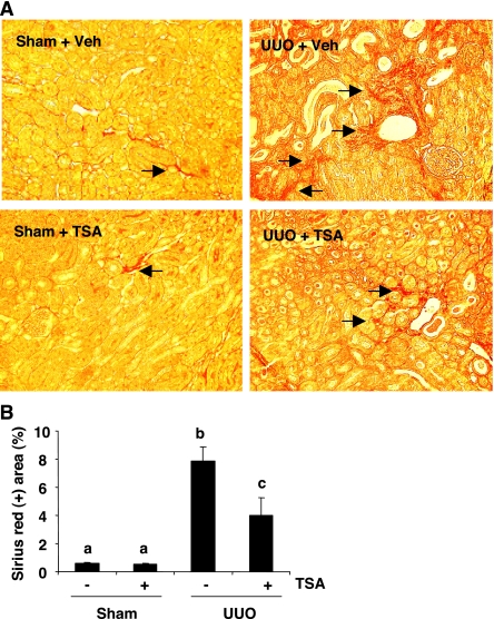
Fig. 7.
Effect of TSA on renal fibrosis after UUO. A: photomicrographs illustrating sirius red staining of sham, UUO-injured kidneys with/without TSA administration. B: graph showing the percentage of sirius red-positive tubulointerstitial area (red) relative to the whole area from 10 random cortical fields (×200). Values are means ± SD. Bars with different letters (a–c) are significantly different from one another (P < 0.05).

