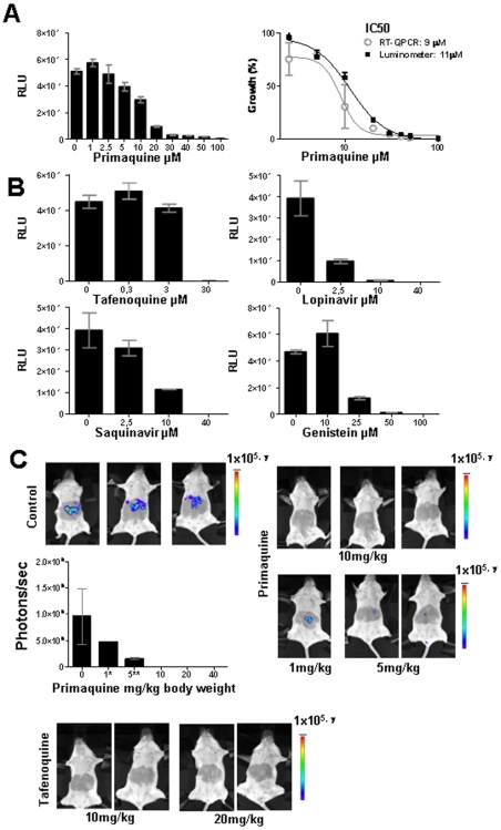
Figure 3. Drug-inhibition of liver stage development determined by measurement of luciferase expression (luminescence).
A. Inhibition of in vitro liver stage development by primaquine (left panel) by measuring luminescence levels (RLU) in samples of Huh7 cells 44 h after infection of the cells with 3×10⁴ PbGFP-Luccon sporozoites. The right panel shows the inhibition of liver stage development by primaquine as determined by both luminescence measurements and qRT-qPCR analysis. The percentage of growth is defined by the RLU values and by the amounts of P. berghei 18S rRNA levels, respectively. Luminescence levels were measured using a Tecan microplate reader. B. Inhibition of in vitro liver stage development by tafenoquine, lopinavir, sanquinavir and genistein, as determined by measuring luciferase luminescence levels (RLU) in samples of Huh7-infected cells 44 h after infection of the cells with 3×10⁴ PbGFP-Luccon sporozoites. Luminescence levels were measured using a microplate reader. C. Inhibition of in vivo liver stage development by primaquine and tafenoquine as determined by measuring luminescence levels (photons/sec) in live mice at 44 h after infection of the mice by the bite of 5 infected mosquitoes. Luminescence levels were determined by direct imaging of whole bodies using the IVIS100 system.

