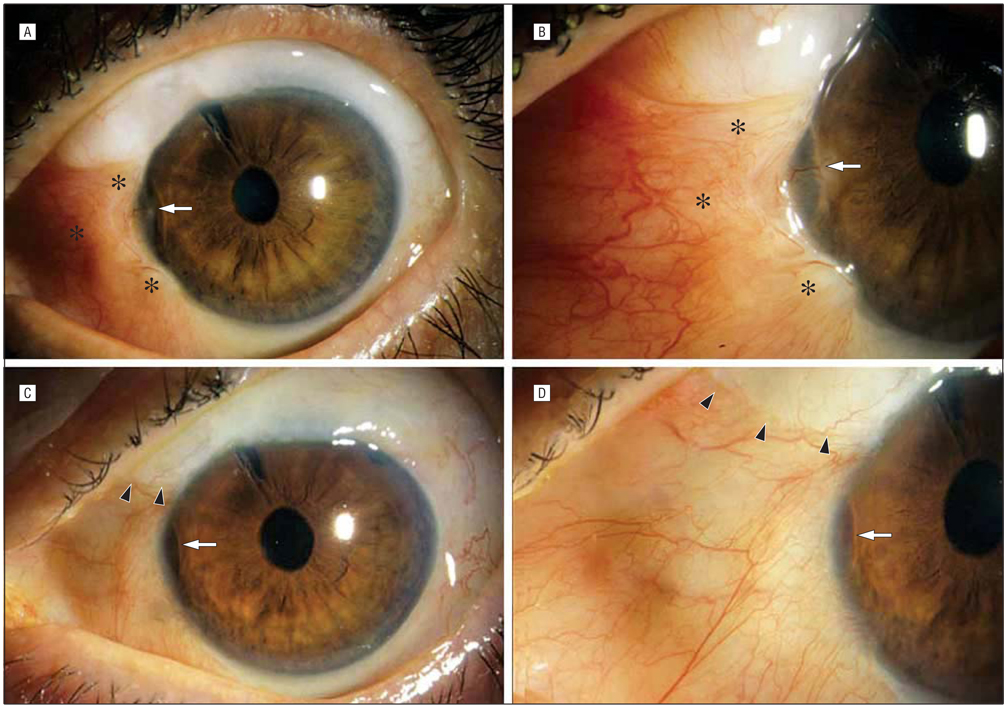
Figure 2.
A, Preoperative photograph of case report patient showing thick vascularized bleb in the nasal interpalpebral space (asterisks) with corneal delle (arrow). B, Close-up showing corneal delle (arrow) with vascularization and thick, elevated bleb extending to limbus (asterisks). C, One-year postoperative photo showing superonasal limit of bleb (arrowheads) with moderate persistence of delle (arrow). D, Close-up postoperative photograph. Comparison to B shows resolution of thick interpalpebral bleb. Delle (arrow) is much improved and no longer vascularized.

