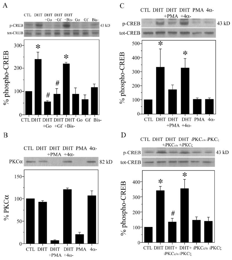
Fig. 4.
PKC contributes to androgen-induced CREB activation in hippocampal neuron cultures. A, Hippocampal neuron cultures were pretreated for 2 h with PKC inhibitor 2 μM GF109203X (GF), Gö6983 (Go), the inactive analog 2 μM bisindolylmaleimide V (Bis-), or vehicle, followed by 2 h exposure to 10 nM DHT, and then assessed by western blot using phosphorylated (p-CREB) and total (tot-CREB) CREB antibodies (43 kDa). Percent phospho-CREB is expressed as a ratio of phosphorylated to total CREB, normalized to the vehicle-treated control condition (bottom panels), [F (7,15) = 6.0; P = 0.002]. B–C, Hippocampal neuron cultures were pretreated with 100 ng/ml PMA, inactive 4α-phorbol (4α-), or vehicle, followed 24 h later by application of 10 nM DHT for 2 h, and then immunoblots were probed for (B) PKCα (82 kDa) and (C) phosphorylated and total CREB [F (5,11) = 2.7; P = 0.075]. D, Neuron cultures were pretreated with 1 μM peptide inhibitors of conventional and novel (iPKCc/n) and atypical (iPKCα) PKC or vehicle for 2 h, and then exposed to 10 nM DHT for 2 h [F (5,11) = 3.6; P = 0.036]. Immunoblots are from representative experiments and data show means (± SEM) of the combined experiments (n = 3). *p < 0.05 relative to the vehicle-treated control condition. # p < relative to the paired DHT condition.

