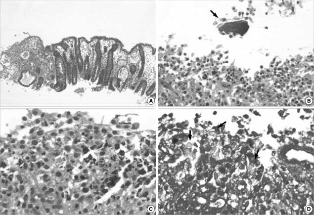
Fig. 3.
Microphotographs of the follow up colonic biopsy. (A) Colonic mucosa shows crypt regeneration and granulation tissue formation (H&E stain, ×40). (B) The remaining Kalimate crystal (arrow) shows a degenerative appearance with an irregular edge and surrounding inflammatory activity (H&E stain, ×200). (C) Small fragments of crystal are embedded in the inflamed granulation tissue (H&E stain, ×400), crystalloid appearances (arrows) of which are highlighted on Diff-Quick stain (D, ×400)

