Figure 2.
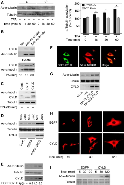
CYLD increases the level of acetylated α-tubulin. (A) Analysis of the levels (left panels) and quantification (right panel) of acetylated α-tubulin and total tubulin in Cyld+/+ and Cyld−/− keratinocytes treated with TPA (15, 30 and 60 min). (B) Immunoprecipitation of endogenous CYLD from keratinocytes treated with 100 nM TPA and immunoblotting against acetylated-α-tubulin or CYLD. The lysate (lower panel) shows equal amount of protein used for immunoprecipitation. (C) Analysis of the levels of acetylated α-tubulin and total tubulin in untreated or TPA-treated (30 min) Cyld−/− keratinocytes (control) or Cyld−/− keratinocytes transiently transfected with full-length-CYLD for 48 h. (D) Analysis of the levels of acetylated α-tubulin, total tubulin, or CYLD in two different melanoma cell lines (MEL Im and Juso) infected with mock, EGFP, or EGFP–CYLD lentivirus (1 × 10⁵ IU) for 24 h. The medium was replaced two times before the cells were lysed and used for immunoblotting against α-tubulin, total tubulin, or CYLD. (E) Analysis of the levels of acetylated α-tubulin, total tubulin, and CYLD in melanoma cells (Juso) transiently transfected with increasing concentrations of EGFP–CYLD cDNA. (F) The confocal plane of human malignant melanoma cell line (Juso) transiently transfected with 1.0 μg EGFP–CYLD (green) for 24 h and stained for acetylated α-tubulin (red). The asterisk indicates cells lacking CYLD expression. (G) Analysis of the levels of acetylated α-tubulin and total tubulin in melanoma cells transduced with 1 × 10⁵ IU of HA, HA–CYLD, or the catalytically inactive mutant HA–CYLDC/S lentivirus for 24 h. The medium was replaced two times before the cells were lysed and used for immunoblotting against α-tubulin, total tubulin, or CYLD. (H) Immunofluorescence staining of microtubule re-growth of EGFP and EGFP–CYLD lentivirus-infected melanoma cells after treatment with nocodazole (50 μM) for 10 min, followed by a washout and incubation in fresh culture medium at 37°C for 10, 30, or 120 min. (I) EGFP or EGFP–CYLD lentivirus-infected melanoma cells were used to analyse the levels of acetylated α-tubulin and total tubulin in untreated cells or cells treated with nocodazole (50 μM) for 10 min followed by a washout and incubation in fresh culture medium at 37°C for 10, 30, or 120 min before cell lysis. *P<0.05.

