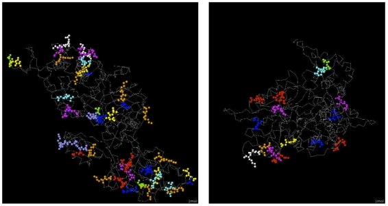
Figure 7. Three dimensional structures of ETF2 and RNR1 proteins obtained from the protein data bank.
Only dimers important for prediction are shown with the rest of the protein structure as backbone. Different colors correspond to different dimers. EE: red, AE: green, AD:blue, DA: yellow, DE: magenta, DV: cyan, EK: white, EQ: violet, KA: orange.

