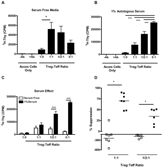
Figure 1. CD4+CD25+ Treg cells require serum for suppression of T cell proliferation.
Suppression assays were conducted utilizing FACS sorted cells run in parallel under either (A) SFM or (B) SFM supplemented with 1.0% autologous serum (n = 7 healthy controls). Treg cells were plated alone (5×10³ cells/well), and in decreasing ratios (1∶1, ½∶1, and 0∶1) to a constant number of Teff cells. Cells were stimulated with soluble anti-CD3 (5.0 µg/ml) and anti-CD28 (2.5 µg/ml) in the presence of a ten-fold excess (5×10⁴) of irradiated accessory cells. (C) Graph indicates data plotted from panels (A) and (B) to highlight the differences in proliferation between serum free media (open bars) and 1% serum (closed bars). Cells proliferated significantly less in SFM at Treg to Teff ratios of ½∶1 and 0∶1 (p<0.001 for both conditions). (D). Graph indicates the percent suppression calculated under serum-free conditions (open squares) and supplemented with 1.0% serum (closed squares) at a ratio of 1∶1-Treg to Teff cells (left data points) and at a ratio of ½∶1 Treg∶Teff cells (right points). Under serum free conditions, Treg fail to suppress proliferation and often lead to increased proliferation in the co-culture (% suppression = mean + SEM, −197.8+270.6 for SFM vs. 70.4+17.3 with 1.0% serum; *p = 0.04 at a ratio 1∶1 Treg to Teff. This trend continued at a ratio of ½∶1 Treg to Teff cell (−103.1+91.5 vs. 34.8+18.12, for SFM and 1.0% serum, respectively; **p = 0.01). (*p<0.05, **p<0.01, and ***p<0.001).

