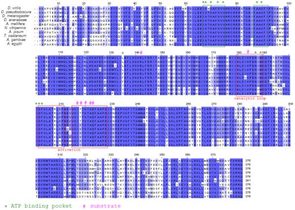
FIGURE 7.

Jalview of CLUSTAL alignments for the kinase domains of projectin. Conserved amino acids are highlighted with different shades of blue to represent the degree of conservation. The position of the ATP and substrate binding sites, as well as the catalytic and activation loops were modeled on this sequence by comparison with twitchin kinase and serine-threonine kinase as available in the Conserved Domain Database (CDD v2.13) at NCBI.
