Figure 3.
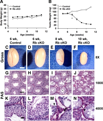
Progressive testicular atrophy and seminiferous tubule breakdown occurs in Rb cKO mice. Comparisons of body weights (A) and testis weights (B) from 5–11 wk of age show that although body weights do not vary significantly between groups, Rb cKO testis weights significantly decrease at 7 wk of age (P < 0.05) and progressively decrease to less than 19% of the control weights by 11 wk of age (n = 6–10 per data point). Gross morphology (C–F) and periodic Schiff (PAS) staining (G–N) of control (C, G, and K) compared with Rb cKO (D–F, H–J, and L–N) testes. A full complement of germ cells can be observed in control (G and K) and Rb cKO (H and L) testes at 6 wk of age, but representative sections from 8 (I and M) and 10-wk-old (J and N) testes show progressive loss of maturing germ cells in addition to reduction of tubular and luminal widths and increased vacuolization. Asterisks (I) indicate tubules in the 8-wk-old Rb cKO testis that had lost elongating spermatids and spermatozoa. Progressive germ cell loss is presumably not completely caused by loss of Sertoli cells because their presence can be identified in these dysfunctional tubules (arrowheads, M and N). C–F, 2 mm; G–J, 100 μm; K–N, 20 μm.

