Figure 6.
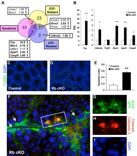
RB deletion in Sertoli cells causes activation of apoptotic pathways. A, Venn diagram of microarray changes in E2F-related, p53-related, and apoptotic genes shows the overlap between these pathways. B, Quantitative RT-PCR of select (bold) genes from panel A, and Trp53 confirms the up-regulation of these genes in Rb cKO Sertoli cells (*, P < 0.05; **, P < 0.01; control, n = 6; Rb cKO, n = 5). RQ, Relative quantification. C–E, TUNEL (green) staining in 6-wk-old testes is increased in Rb cKO (D) as compared with control (C) as determined by apoptotic index (E) (**, P < 0.01; control, n = 3; Rb cKO, n = 4). F–I, Immunofluorescent analysis of cleaved caspase-3 (red) and Sox9-GFP (green) reveals the activation of caspase-mediated apoptosis in 6-wk-old Rb cKO Sertoli cells. Arrows (I) indicate Sertoli cells that did not stain for cleaved caspase-3. DAPI, 4′,6-diamidino-2-phenylindole.

