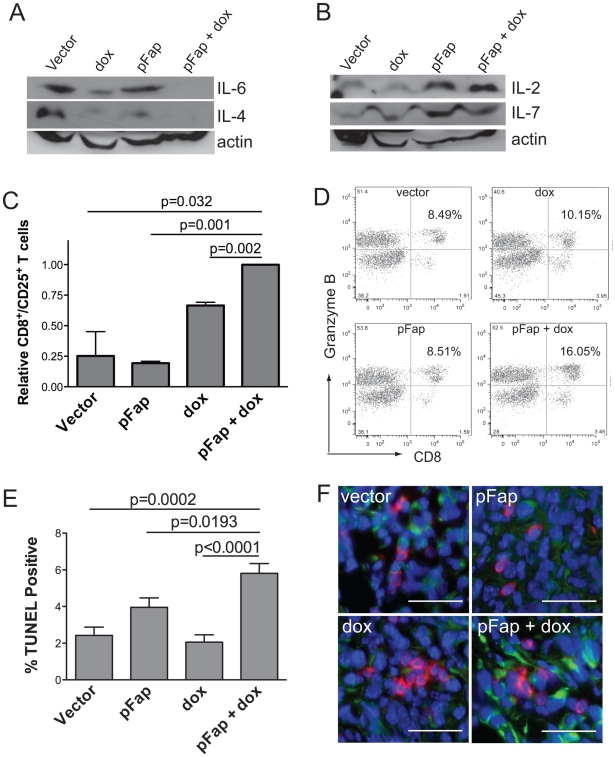
Figure 2. Vaccination with pFap results in Th2 to Th1 cytokine transition in the tumor microenvironment.
Mice were treated in a prophylactic setting and primary tumors isolated 25 days later. (A–B) Tumor homogenates were analyzed by Western blotting to determine IL-6 and IL4 (A) and IL-2 and IL-7 (B) protein levels. (C) Live primary tumor cell suspensions were analyzed by flow cytometry using anti-CD8 and anti-CD25 antibodies to detect activated T-cells. Results are shown as percent of CD8+/CD25+ T cells relative to mice treated with the combination therapy. (D) Splenocytes were isolated from tumor bearing mice treated with the combination therapy or controls. Splenocytes were stimulated twice with IL-2 and then incubated with irradiated 4T1 tumor cells for 24 hours prior to detection of CD8+/Granzyme B+ cells by flow cytometry. (E) Primary tumors were isolated from mice treated in a prophylactic setting 10-days after the final treatment with doxorubicin and apoptotic tumor cells were visualized by quantified using the TUNEL assay. (F) Additionally, we also used immunohistochemistry to visualize CD8+ T cells (red) and apoptotic tumor cells expressing caspase 3 (green) in primary tumors. Scale bar = 100 µm.

