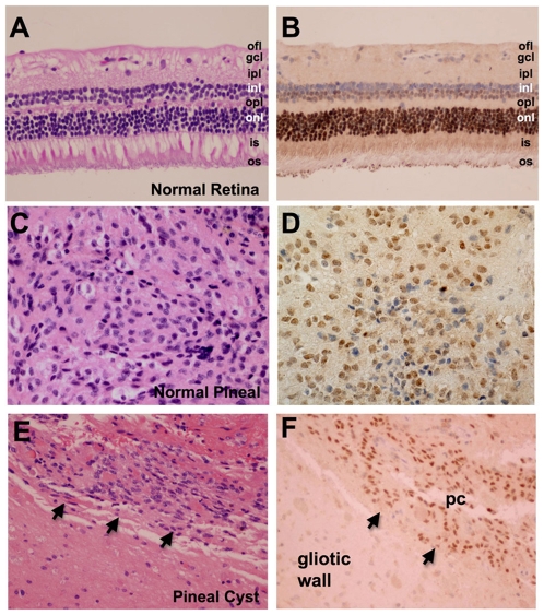
Figure 3. CRX protein is highly expressed in the normal adult human retina and pineal.
Immunohistochemical analysis of CRX expression demonstrates robust expression in the nuclei of cones and rod cells in the outer nuclear layer (onl) as well as much weaker expression in a subset of nuclei in the inner nuclear layer (inl) that abut the outer plexiform layer (opl) of the adult human retina (A, B). This region is populated predominantly by horizontal and bipolar cells. The ganglion cell layer does not express CRX. Expression of CRX in the pinealocytes of a surgically resected normal pineal gland (C, D) and pineal cyst (E, F arrows). Gliotic regions composing the cyst wall do not express CRX (lower left corner of E, F). Optic fiber layer (ofl), ganglion cell layer (gcl), inner plexiform layer (ipl), inner segment (is), outer segment (os), pineal cyst (pc). Original magnification 200x (A, B, E, F) and 400x (C, D).

