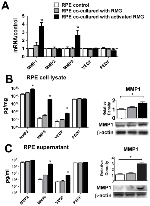
Figure 5. Primary RPE cells increase expression and secretion of pro-angiogenic factors following co-culture with retinal microglia (RMG).
(A) Quantitative RT-PCR comparing mRNA levels of metalloproteinases (MMP1, MMP2, MMP9), and growth factors, VEGF and PEDF, in RPE cells co-cultured with unactivated (gray bars) and activated RMG (black bars) relative to control (unexposed RPE cells, white bars). (B) Protein analyses of RPE cell lysates showing that levels of MMP2, MMP9, and VEGF, as analyzed by ELISA, were increased following RMG co-culture (left). MMP1 levels, as analyzed by Western blotting, were also significantly elevated (right). (C) Protein analyses of RPE supernatants showing similar significant increases by ELISA analyses (MMP9, VEGF, left) and by Western blotting (MMP1, right). Levels of PEDF, an anti-angiogenic factor, were unchanged by co-culture in both RPE cell lysates and supernatants. Significant differences (p<0.05) are indicated with *.

