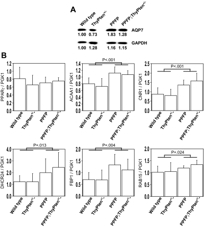
Figure 4.
Gene expression in PPFP mice. Thyroid glands were analyzed for the expression of genes previously shown to be induced in human PPFP thyroid cancer. A, Western blot analysis of AQP7 showing increased expression in PPFP mice. The blot was reprobed for GAPDH as a loading control. The numbers below each band indicate band intensities normalized to wild-type AQP7 (upper immunoblot) or wild-type GAPDH (lower immunoblot). Each lane contains 40 μg protein pooled from four mice. B, Real-time RT-PCR analysis of gene expression normalized to PGK1. The bars show the means ± sd for five mice. Statistical analyses were performed by one-tailed t test comparing the 10 mice expressing PPFP (PPFP and PPFP;ThyPten+/−) with the 10 mice without PPFP (wild-type and ThyPten+/−).

