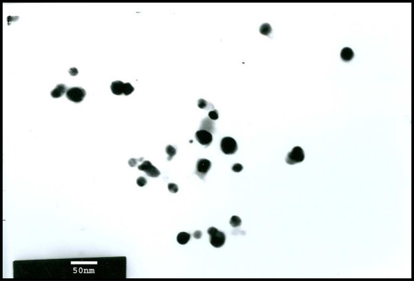
Figure 1.
TEM images obtained from purified fractions collected after sucrose density gradient of Ag-NPs synthesized using B. licheniformis. Purified nanoparticles from B. licheniformis were examined by electron microscopy. Several fields were photographed and were used to determine the diameter of nanoparticles. The range of observed diameters is 50 nm.

