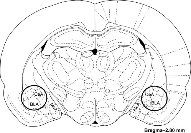
Figure 1. Schematic image depicting the location of the amygdala tissue punches in experiment 2.
The whole rat brain was placed in a block and it was cut into 2-mm-thick coronal sections. The majority of the amygdala was removed with a 2 mm diameter punch tool from the tissue slice that was located from –1.6 to –3.6 mm posterior of bregma. The punches are depicted as dark solid line circles on the image adapted from an atlas (Paxinos & Watson 1998).

