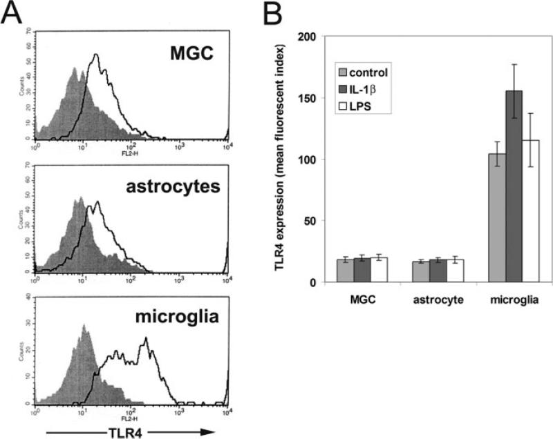
Fig. 3.
TLR4 expression by mixed glial cultures, pure astrocytes, and pure microglia. Mixed glial cultures (MGC) and pure cultures of astrocytes or microglia were prepared as described in the Materials and Methods section, switched to serum-free conditions for two days, and then analysed for cell surface expression of TLR4 (isotype control antibody: shaded filled line; TLR4 antibody: open line). These expression profiles are representative of three different experiments performed. Note that TLR4 was expressed at a similar low level by mixed glial cultures and pure astrocytes, but at a much higher level by microglia (A). The influence of IL-1β or LPS on TLR4 expression in astrocytes and microglia was investigated by incubating the three different types of culture with IL-1β (10 ng/ml) or LPS (20 ng/ml) for 2 days, before analyzing TLR4 expression by flow cytometry (B). The points represent the mean ± SD of three experiments. Note that mixed glial cultures and pure astrocytes showed the same low level of TLR4 expression, which was not affected by IL-1β or LPS. In contrast, microglia express markedly higher levels of TLR4 and this was significantly increased by IL-1β.

