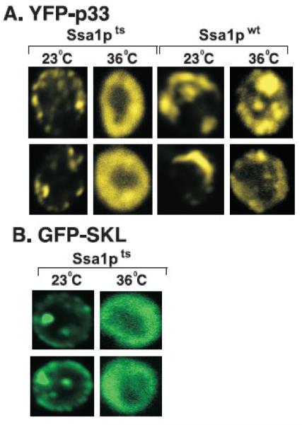
Fig. 3.
Cytosolic localization of p33 replication protein in ssa1ts yeast grown at 36°C. (A) Confocal laser microscopy shows the localization of YFP-p33 in ssa1ts yeast and SSA1wt yeast grown at either 23°C or 36°C for 14 hours after induction from GAL1 promoter. The punctate structures show peroxisomal localization, while the “doughnut” -shape structure indicates cytosolic localization of p33 in yeast cells. The punctate structures formed in ssa1ts yeast grown at 23°C are reminiscent of those found in SSA1wt yeast (left side of the panel). (B) Cytosolic localization of peroxisome-targeted GFP, termed GFP-SKL, in ssa1ts yeast grown at 36°C for 14 hours after induction from GAL1 promoter. Note that GFP-SKL carries the peroxisomal targeting sequence “SKL” at the C-terminus.

