
FIGURE 3.
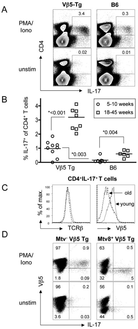
Mtv-8+ Vβ5 Tg mice have elevated frequencies of Th17 T cells that display revised TCR. Splenocytes from 5- to 10-wk-old and 18- to 45-wk-old B6 and Vβ5 Tg mice were stimulated for 6 h with PMA/Ionomycin (PMA/Iono) or left untreated (unstim). A, Representative flow data are depicted for total splenocytes from 23-wk-old mice. Percentages refer to IL-17+ T cells among total CD4+ T cells. B, For all analyzed mice, the fraction of CD4+ T cells producing IL-17 in response to PMA/ionomycin stimulation is plotted and the p values determined by Student's t test are shown. C, The levels of pan-TCRβ (left panel) or Vβ5 staining (right panel) are depicted for gated CD4+IL-17+ cells from representative young and old Vβ5 Tg mice. D, Splenocytes from a 40-wk-old Mtv− Vβ5 Tg (left panels) and a Mtv-8+ Vβ5 Tg mouse (right panels) were stimulated for 6 h with PMA/Ionomycin (upper row) or left untreated (lower row). Shown are CD4+ gated T cells stained for surface Vβ5 and intracellular IL-17.