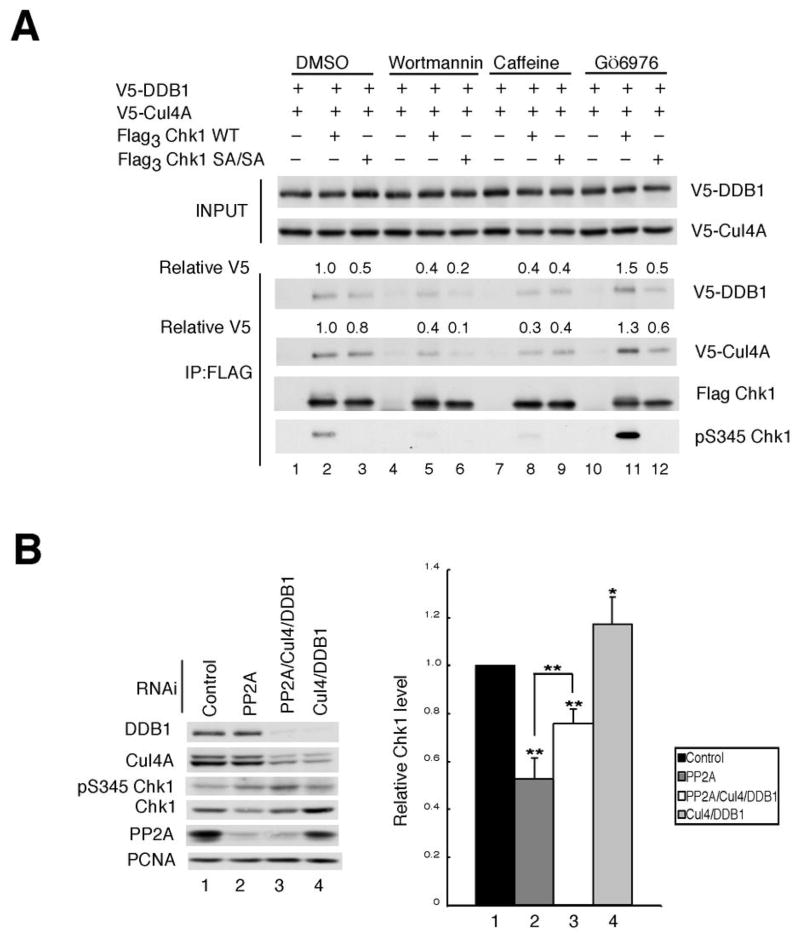
Figure 2.
Chk1 phosphorylation regulates interactions with Cul4A and DDB1. A, asynchronously growing HeLa cells expressing the indicated tagged proteins were cultured in the presence of vehicle (DMSO), 50 μM wortmannin, 10 mM caffeine or 1 μM Gö6976 for 2 h each. Cells were then exposed to 20 mM HU and 10 μM MG132 for 4 h prior to harvest. Flag3 Chk1 was immunoprecipitated and precipitates were analyzed by Western blotting. Relative levels of DDB1 and Cul4 are indicated (n = 3). B, asynchronously growing HeLa cells were treated with control RNAi or RNAi specific for PP2A, Cul4A/DDB1 or PP2A/Cul4A/DDB1 for 48 h. Lysates were prepared and immunoblotted with antibodies specific for the indicated antibodies (left panel). The data from 4 independent experiments is presented graphically in the right panel. The data is presented as mean +/− standard deviation. P-values from Student’s t-test are shown when significantly different from control. Asterisks indicate significant p-values (* < 0.05; **<0.01)

