
Figure 5.
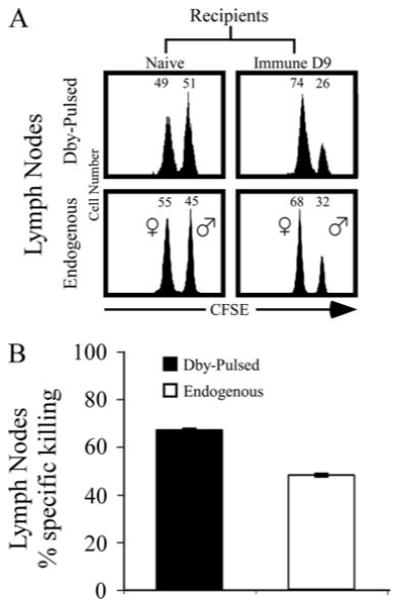
MHC class II-restricted cytolytic activity following immunization with male cells is detectable 9 days postimmunization. A, Top panels, equal numbers of Ly5.1 CFSEhigh Dby peptide-pulsed and CFSElow control peptide-pulsed female splenocytes were injected i.v. into Ly5.2 naive or day 9 immune female mice. In vivo cytotoxicity was assessed in the lymph nodes 48 h after transfer. Representative histograms are gated on Ly5.1+MHC class IIhigh donor cells. Numbers at the top of each plot represent the percentage of CFSElow or CFSEhigh cells of the total Ly5.1+MHC class IIhigh donor cells recovered. A, Lower panels, equal numbers of Ly5.1 β2m−/− CFSEhigh male and CFSElow female splenocytes were coinjected into Ly5.2 naive or day 9 immune NK-depleted female mice. In vivo cytotoxicity was assessed in the lymph nodes 48 h after transfer. Representative histograms are gated on Ly5.1+MHC class IIhigh donor cells. Numbers at the top of each plot represent the percentage of CFSElow or CFSEhigh cells of the total Ly5.1+MHC class IIhigh donor cells recovered. B, Cumulative data from three independent experiments of three mice per group. Error bars, SEM.