
FIGURE 1. SDS-PAGE analysis and autophosphorylation assays of RegB.
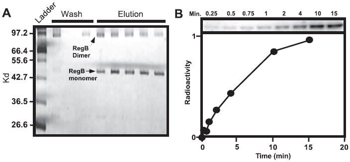
A, solubilized and purified RegB separated by SDS-PAGE. Lane 1 contains the molecular weight mass followed by the wash fraction and the elution fraction after the addition of imidazole. B, autophosphorylation of purified monomer RegB using γ-32P-labeled ATP as a tracer. The aliquots were removed at 0.25, 0.5, 0.75, 1, 2, 4, 10, and 15 min after the addition of ATP, and the reaction was quenched with SDS-PAGE loading buffer before being separated by SDS-PAGE. The graph below the autoradiograph represents the arbitrary units of 32P incorporation derived from phosphorimaging data analysis.