
FIGURE 4. Topological representation and homolog pile up of the transmembrane-spanning domains of RegB.
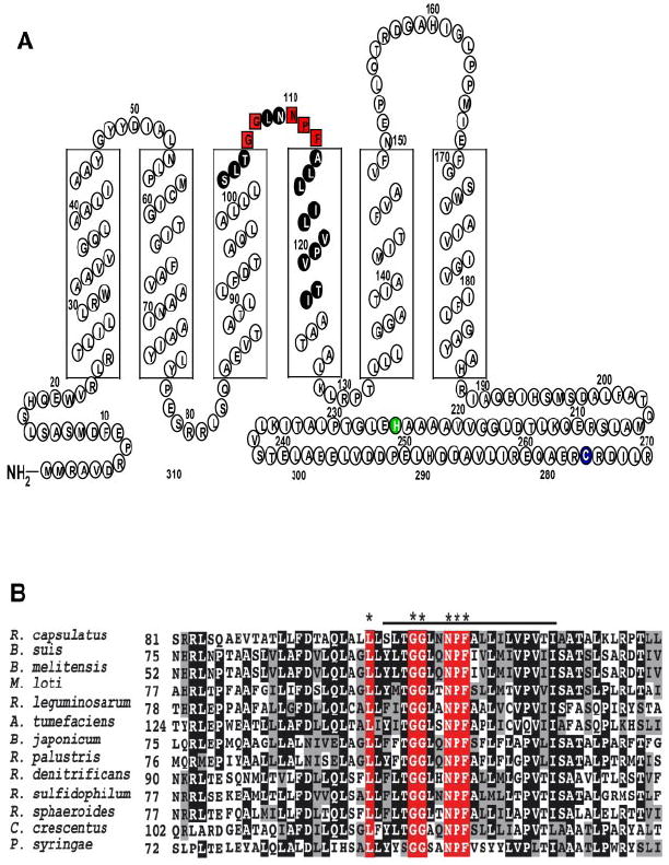
A, RegB has six transmembrane-spanning domains that anchor it to the bacterial membrane. The boxed red letters represent the 100% conserved pentapeptide ubiquinone-binding motif. The amino acids of the peptide fragment identified by HPLC and azido-Q cross-linking experiments are colored black. The site of phosphorylation is colored green, and the redox active Cys265 is colored blue. Amino acid residues past 270 were omitted for simplicity. B, RegB homologs were selected from Brucella suis, Brucella melitensis, Mesorhizobium loti, Rhizobium leguminosarum, Agrobacterium tumefaciens, Bradyrhizobium japonicum, Rhodopseudomonas palustris, Roseobacter denitrificans, Rhodovulum sulfidophilum, R. sphaeroides, R. capsulatus, Caulobacter crescentus, and Pseudomonas syringae. The asterisks denote the 100% conserved residues in the newly identified ubiquinone-binding site. The alignment was created using ClustalW and the ALN file was imported into BoxShade to color code the conserved residues in black, similar residues in gray, and nonconserved residues in white. The line above the alignment denotes the identified ubiquinone-binding peptide.