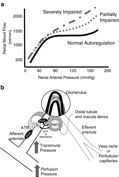
Fig. 3.
Postulated signalling mechanisms for ATP-mediated autoregulatory adjustments in afferent arteriolar diameter. Panel a illustrates a normal profile for autoregulation of renal blood flow (solid black line) and the autoregulatory profiles that might be observed under conditions of moderate and severe autoregulatory dysfunction (modified from [171]). b ATP might be released from afferent arteriole smooth muscle cells in response to an increase in renal perfusion pressure. This ATP released into the perivascular interstitial fluid could act upon P2X1 receptors to evoke autoregulatory vasoconstrictor responses. ATP released from the macula densa, in response to an increase in distal tubular fluid NaCl concentration, can traverse the interstitial fluid of the juxtaglomerular apparatus to act on afferent arterioles and induce tubuloglomerular feedback-mediated vasoconstriction. Alternatively, released ATP could be degraded to adenosine in the interstitial fluid prior to activating afferent arteriolar adenosine A1 receptors to regulate afferent arteriolar resistance

