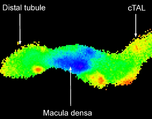
Fig. 3.
Wide-field image of an isolated perfused thick ascending limb. The glomerulus is not shown because fura-2, the calcium-sensitive fluorophore, was loaded through the lumen. High levels of cytosolic calcium are red and yellow, while lower values are green and the lowest calcium concentrations are in blue. The macula densa cells have a much lower cytosolic calcium concentration compared to the surrounding epithelial cells

