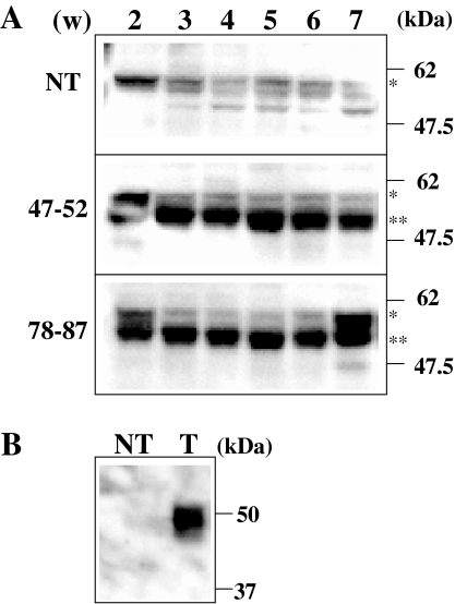
Fig. 2.
Western blot analysis of rice seed proteins. a Soluble proteins were extracted from seeds after 2–7 weeks (w) of self-pollination. Total proteins (10 μg) were loaded on 10% SDS–PAGE, followed by electroblotting. Primary and secondary antibodies used were anti-GAD2 antibody and anti-rabbit IgG antibody conjugated with alkaline phosphatase (SIGMA), respectively. Positions of protein marker are shown on the right side. A single and double asterisks are corresponded to protein bands of GAD2 (57 kDa) and GAD2ΔC (53 kDa), respectively. b Control experiment of western blot analysis. NT: calli from non-transformed rice, T: calli over-expressing GAD2ΔC described in Akama and Takaiwa (2007)

