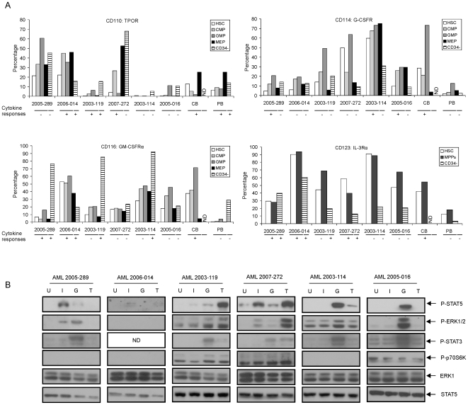
Figure 3. Cytokine and growth factor receptor expression in normal and leukemic cells.
The expression of receptors for TPO (CD110), GM-CSF (CD116), IL-3 (CD123) and G-CSF (CD114) was analyzed by FACS. (A) MNCs from AML samples and CD34+ cells CB and PB were stained with antibodies against receptors, together with CD34, CD38, CD45RA and CD123 for 30 minutes. CD123 expression was analyzed in HSCs, MPPs (CD34+CD38+) and CD34− populations. For CB cells, no CD34− fraction was available. The cytokine responses for STAT5 activation in CD34+ versus CD34− populations are also indicated (“+” indicates that STAT5 was activated by the respective cytokine; “−” indicates that STAT5 was not activated). (B) 1×10⁶/ml CD34+ cells were sorted by MoFlo from AML MNCs and suspended in HPGM for 2 hours. Cells were harvested after stimulation with 1.0 ng/ml IL-3 (I), 100 ng/ml G-CSF (G) or 100 ng/ml TPO (T) for 15 minutes or left unstimulated (indicated as U) for Western bloting. ND, not determined.

