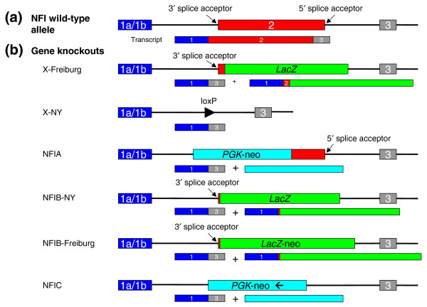
Figure 1.
For simplicity the same structure is drawn for all four NFI genes. (a) The organization of the NFI genes. They can all use an alternative exon 1, here denoted as a single box labeled 1a/1b. The DNA-binding and dimerization domains are located in exon 2. (b) In general, two approaches are used for knockouts of these genes. The first relies on complete deletion of the second exon (including 5' and 3' splice acceptor sites of proximal introns), as shown here in the X-NY knockout. The second strategy is to insert LacZ (or a LacZ-neo hybrid or PGK-neo hybrid) in-frame into the second exon, leading to production of a fusion protein composed of a few amino acids derived from exons 1 and 2 of the NFI and LacZ genes. In all cases an alternative splice variant joining the first and the third exon of the NFI gene will be formed. The third exon is not in frame with the first, and so premature termination of translation will occur. Whether a peptide produced from the joining of exons 1 and 3 has any physiological function was never analyzed, but judging from the very different phenotypes of the different knockout strains it seems rather unlikely. The NFIB constructs are reported in [15,16], the NFIA knockout in [17] and the NFIC knockout in [18].

