Fig. 3.
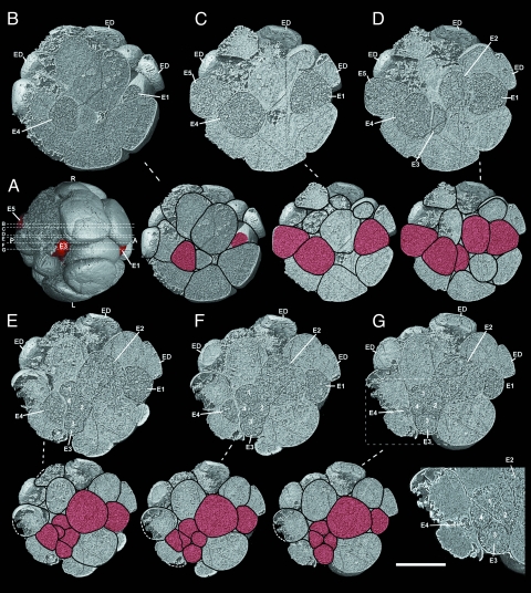
Precambrian postgastrular embryo (4F4) from the Weng'an Phosphate Member of the Doushantou Formation as serial digital sections that cut in at successively deeper layers from the right surface, indicated by the lines B–G both in Fig. 3A and Fig. 4 D and E, and the same sections with drawings of cell boundaries. (A) Digital external view from ventral side of the embryo 4F4, showing: E5, E3, and E1 cells (in red) and the positions of digital internal sections in Fig. 3 B–G indicated by lines B–G. (B) Internal section and the same section with drawing of cell boundaries at reduced size cut in 29% from right surface, showing principal endodermal cells E4 and E1 sectioned near the right margin of the endodermal cord. (C) 35% from right surface, showing endodermal cells E5, E4, and E1. (D) 43% from right surface. (E) 46% from right surface. (F) 51% from right surface. (G) 53% from right surface, showing most parts of the endodermal cord, close to the middle plane of the embryo, and cluster of four cells, apparently the daughter cells resulting from mitosis of E3 (enlarged in lower right panel). Key: A, anterior; P, posterior; D, dorsal; V, ventral; L, left side; R, right side; and ED, ectodermal cell. In reduced sections with cell boundaries drawn endodermal cells are in red. (Scale bar: 250 μm in digital sections, 375 μm in the same section with drawing of cell boundaries, 181 μm in the lower right panel.)

