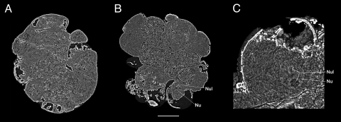
Fig. 5.
Preservation of yolk granules and possible nucleus. (A) Internal section cut inward 39% from dorsal surface, showing detail of the yolk-rich, internal cells in embryo 4F10. (B) A digital thin slice (0.1-μm thick) of an internal section of embryo 4F4 46% from right surface (rotated, but same plane of section as in Fig. 3E), revealing the yolk-rich endodermal cells, as well as the location of a putative nucleus and nucleolus. (C) Internal section of Fig. 5B cell showing a close-up of the putative nucleus and nucleolus, as seen in Movie S3. Key: Nu, nucleus; Nul, nucleolus. (Scale bar, 142 μm in A, 156 μm in B, and 49 μm in C.)

