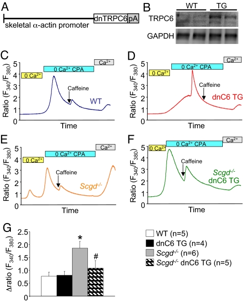
Fig. 4.
SOCE in dystrophic fibers is reduced by inhibition of TRPC activity (A) schematic representation of the dnTRPC6 transgene. (B) Western blot for expression of the dnTRPC6 protein in quadriceps from WT and TG mice. (C) Representative fura-2 tracings from a single WT FDB myofiber subjected to the buffer conditions shown while simultaneously measuring fluorescence. (D–F) Representative fura-2 tracings from a single dnTRPC6, Scgd−/−, or double Scgd−/− dnTRPC6 FDB myofiber subjected to the buffer conditions shown while simultaneously measuring fluorescence (G) Quantitation of multiple tracings from the indicated groups for SOCE as a change in the fura-2 fluorescence ratio. The number of mice used is shown, and 5–8 fibers were analyzed per mouse. *, P < 0.05 vs. Wt and dnTRPC6 Tg. #P < 0.05 vs. TRPC3 TG.

