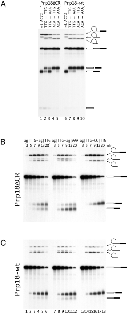
Fig. 3.
Analysis of alternative 3′ splicing in vitro. (A) Splicing of four substrates with alternative 3′ splice sites and wt ACT1 was assayed using extract from a PRP18-knockout strain supplemented with either Prp18ΔCR or Prp18-wt protein. The sequences of the three exon bases at each 3′ splice site are shown at the top; the substrates were based on AS1 (Fig. 1A). The positions of the pre-mRNA, lariat-intron and exon1 intermediates, released lariats (long and short), and alternatively spliced mRNAs are indicated. Splicing was carried out for 15′. (B and C). Kinetics of splicing of three substrates with (B) Prp18ΔCR and (C) Prp18-wt proteins. The 3′ splice sites of the AS1-based substrates are shown at the top of B. In the substrate in lanes 13–18, the downstream AG was mutated to CC. The positions of the RNAs are indicated.

