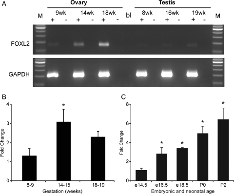
Figure 1.
Detection and quantification of FOXL2 gene expression in the human fetal gonad. (A) Expression of FOXL2 and GAPDH mRNA in the human fetal ovary and testis. Samples at indicated gestational age were analysed by RT–PCR. ‘+’ and ‘−’indicate the presence or absence of reverse transcriptase during cDNA synthesis. M: 100 bp ladder, bl: water blank in PCR reaction. (B) Quantitative PCR measurement of FOXL2 expression in the human fetal ovary at gestations of 8–9 weeks, 14–15 and 18–19 weeks as indicated, n = 4–5 per group. (C) Quantitative PCR measurement of Foxl2 expression in embryonic and post-natal mouse ovary at ages indicated, n = 3-5 per group. Both human and mouse data were analysed by ANOVA with Duncan's post hoc test using earliest gestation as comparator, *P < 0.05.

