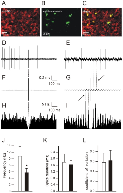
Figure 4. Golgi cells in BK−/− mice exhibit rhythmic firing phase-locked with the LFPO.
(A–C) Representative confocal laser scan microscopy images of mouse cerebellar cortex granular layer containing numerous and tiny granule cells, a bit larger unipolar brush cells and the much larger Golgi cells. The immunofluorescence of the granular layer shows BK channel expression (A; red) in tiny granule cells and in larger neurons as well as somatostatin expression, a marker for Golgi cells, in a fraction of large neurons (B; green). The colocalization (yellow) demonstrates BK channel-positive Golgi cells (C); scale bars: 25 µm. (D–I). Extracellular recording of a Golgi cell in a WT (D) and a BK−/− mouse (E) with spike triggered averaging (F–G) and autocorrelograms (H–I) of the corresponding recordings. Note the phase-locking and the rhythmicity of Golgi cells in BK−/− mice. (J–K) Bar graphs of Golgi spike frequency (J), duration (K), and CV (L) in WT (n = 5) and in BK−/− mice (n = 5).

