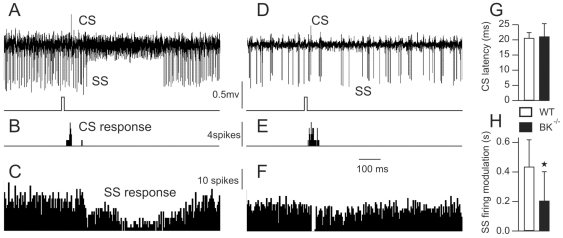
Figure 6. Simple spike response of Purkinje cells to tactile stimulation is altered in BK−/− mice.
(A) Purkinje cells recorded in the Crus2A of a WT mouse during the stimulation of the whisker region, timing of stimulation is illustrated in the lower trace (CS = complex spike, SS = simple spike). (B,C) Bar graphs of complex (B) and simple (C) spike firing, 15 trials summed. (D) Purkinje cells recorded in the Crus2A of a BK−/− mouse during the stimulation of the whisker region. (E,F) Bar graph of complex (E) and simple (F) spike firing, 26 trials summed. (G) Bar graph of timing between stimulus and complex spike firing in WT and BK−/− mice. (H) Bar graph of simple spike response duration in WT and BK−/− mice.

