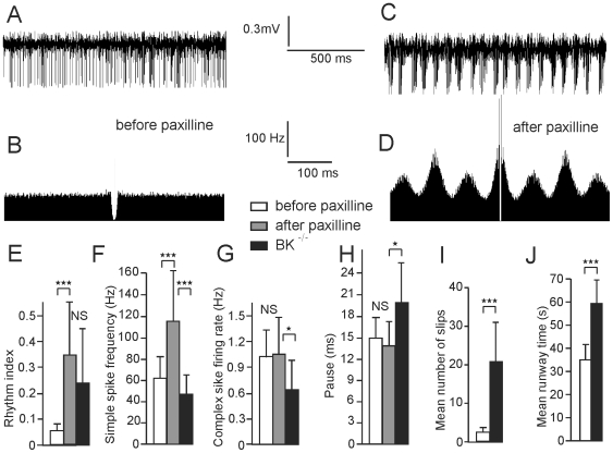
Figure 7. Intracerebellar microinjection of paxilline in WT mice reproduces the rhythmic firing of Purkinje cells and the ataxic behavior of BK−/− mice.
(A,B) Spontaneous firing of a Purkinje cell recorded in a WT mouse (A) and corresponding autocorrelogram (B). Note the absence of rhythmicity. (C,D) The same, following microinjection of paxilline. (E–H) Bar graphs of Purkinje cells simple spike rhythmicity (n = 13)(E) and frequency (n = 13)(F), Purkinje cells complex spike frequency (n = 8)(G) and subsequent pause duration (n = 8) (H) before and after paxilline injection and in BK−/− (n = 48, value illustrated in fig 2 and reproduced here for comparison purpose). Stars indicate significance as in fig 2, for student t test for paired values (comparison between before and after injection) and unpaired values (comparison between WT PC after injection and PC in BK−/−) (I,J) Runway test, bar graph of mean number of slips (I) and time to reach the end of the bar (J) before and after paxilline injection (n = 9).

