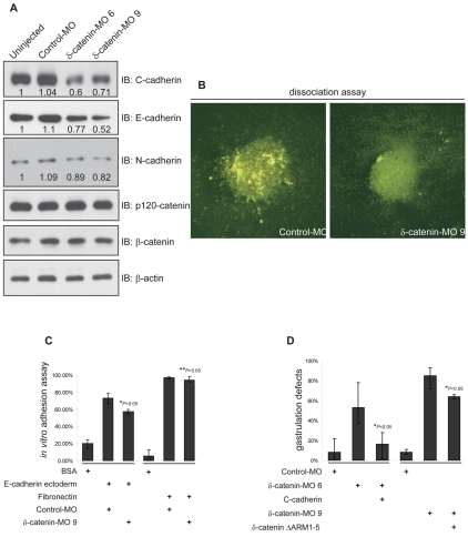
Fig. 7.
δ-catenin depletion leads to reduced cadherin functions. (A) Immunoblotting shows that δ-catenin depletion reproducibly leads to reduced C-, E- and N-cadherin levels, whereas p120-catenin and β-catenin levels are not significantly altered.(B) Calcium-dependant adhesive functions were decreased in δ-catenin-depleted naive ectoderm cells. Following calcium removal from ectoderm explants, note the larger cell aggregates remaining after control MO injection. (C) Using an in vitro assay with the extracellular domain of E-cadherin tethered to a solid substrate (chamber glass), cadherin-mediated adhesion is decreased in naive ectoderm cells depleted of δ-catenin. By contrast, a similar assay that uses tethered fibronectin, did not resolve changes in cell attachment (presumably integrin mediated). (D) A titrated dose of exogenous C-cadherin significantly rescues blastopore closure defects induced by depletion of endogenous δ-catenin. By contrast, a δ-catenin mutant construct lacking armadillo repeats 1-5, and failing to co-immunoprecipitate with C-cadherin (supplementary material Fig. 5A) showed minimal rescuing effects. For all panels P-values indicate statistical significance.

