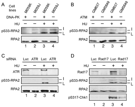
Fig. 3.
S33-RPA2 phosphorylated by ATR in response to replication stress. (A,B) The M059J (DNA-PK–/–) or M059K (DNA-PK+/+) paired cell lines (A), and GM637 (ATM+/+) or GM5849 (ATM–/–) lines (B) were either mock-treated or treated with 2.5 mM HU for 3 hours to cause replication stress. (C) U2-OS cells were transfected with an siRNA directed against ATR or a control siRNA against luciferase (Luc). After 48 hours, cells were either mock-treated or treated with 2.5 mM HU for 3 hours. (D) U2-OS cells were transfected with an siRNA directed against Rad17 or luciferase. For all panels, cells were lysed following treatment and the lysates subjected to SDS-PAGE and western blotting using antibodies specific to total RPA2, S33-P-RPA2, ATR, Rad17 or S317-P-Chk1, as indicated. The total RPA2 signal was used as a loading control. The positions of the various RPA2 species are indicated as in the Fig. 1 legend.

