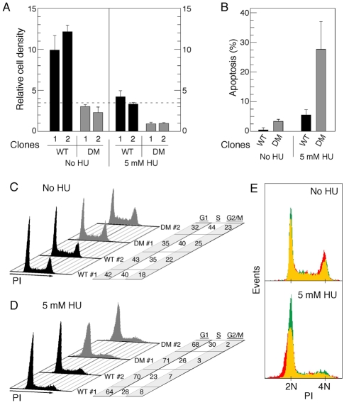
Fig. 8.
Mutation of RPA2 PIKK sites impairs cell growth and alters the cell-cycle profile. (A) Two U2-OS clones each were replaced with either wt- or T21A/S33A-RPA2 (DM, double mutant). At 72 hours post-siRNA transfection, cells were plated in triplicate to an equivalent relative density (0.35; indicated by dashed line) and then either mock-treated or incubated with 5 mM HU. 48 hours later, cells were released from the plate by trypsin-EDTA treatment and cell count in each plate determined. (B) U2-OS clones replaced with either wt- or T21A/S33A-RPA2 (DM) were, at 72 hours post-siRNA transfection, mock-treated or incubated with 5 mM HU for 24 hours. Cells were then processed to quantify the percentage of cells undergoing apoptosis, using a TUNEL assay. (C,D) Similar aliquots of cells described in A were either mock- or HU-treated (5 mM for 48 hours) and then subjected to FACS analysis for DNA content. Cell-cycle profiles and relative distributions (in percent of total) in the G1, S and G2-M phases are shown. (E) Overlay of representative cell distributions from clones replaced with wt-RPA2 (green) or T21A/S33A-RPA2 (red), prepared as described in B and C. Regions of overlap are shown by yellow coloring.

